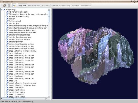
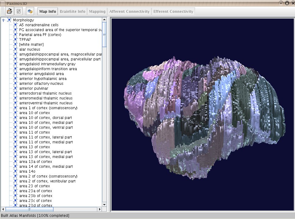
A 3D atlas of brain structures from sections
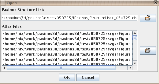
The atlas is built from 151 Scalable Vector Graphics files (SVG) exported from polygon drawings of brain structures. Each file represents a section, page, within the atlas. Andrew Janke has aligned the files using some tools from the medical imaging community. The files are names 'Figure 001.svg', most anterior section, to 'Figure 151.svg', most posterior section. The standard distance between sections is 0.45 mm, though deviations exist between the first (1-8) and last (140-151) sections.Within each file the brain structures are represented by SVG paths. Each structure has a unique fill colour indicated in hex format. An Excel file stores the list of structures and corresponding RGB colours (in decimal format).
Almost all drawn brain structures are referenced to our CoCoMac connectivity database so that we can later extract relevant information from the database with the aim to display it on the atlas.
Paxinos3D is available for redistribution under these terms of the Lesser General Public License, LGPL.
Documentation
Downloads
|
|
|
||||
|
|
Download the Paxinos3D source files. |
[NOTE: Paxinos3D is currently only available for Linux and Windows. Other platforms such as MacOS X can be supported if required.]
please email all feedback to nix